Abstract
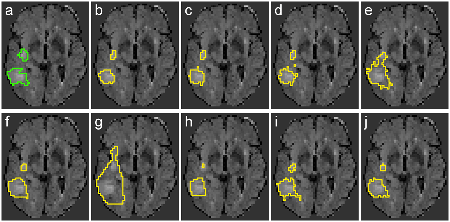
Motivation Ischemic stroke, triggered by an obstruction in the cerebral blood supply, leads to infarction of the affected brain tissue. An accurate and reproducible automatic segmentation is of high interest, since the lesion volume is an important end-point for clinical trials. However, various factors, such as the high variance in lesion shape, location and appearance, render it a difficult task. Methods In this article, nine classification methods (e.g. Generalized Linear Models, Random Decision Forests and Convolutional Neural Networks) are evaluated and compared with each other using 37 multiparametric MRI datasets of ischemic stroke patients in the sub-acute phase in terms of their accuracy and reliability for ischemic stroke lesion segmentation. Within this context, a multi-spectral classification approach is compared against mono-spectral classification performance using only FLAIR MRI datasets and two sets of expert segmentations are used for inter-observer agreement evaluation. Results and Conclusion The results of this study reveal that high-level machine learning methods lead to significantly better segmentation results compared to the rather simple classification methods, pointing towards a difficult non-linear problem. The overall best segmentation results were achieved by a Random Decision Forest and a Convolutional Neural Networks classification approach, even outperforming all previously published results. However, none of the methods tested in this work are capable of achieving results in the range of the human observer agreement and the automatic ischemic stroke lesion segmentation remains a complicated problem that needs to be explored in more detail to improve the segmentation results.
BibTeX
@article{Schroeder2015StrokeLesionSegmentation,
 abstract = {Motivation Ischemic stroke, triggered by an obstruction in the cerebral blood supply, leads to infarction of the affected brain tissue. An accurate and reproducible automatic segmentation is of high interest, since the lesion volume is an important end-point for clinical trials. However, various factors, such as the high variance in lesion shape, location and appearance, render it a difficult task.   Methods In this article, nine classification methods (e.g. Generalized Linear Models, Random Decision Forests and Convolutional Neural Networks) are evaluated and compared with each other using 37 multiparametric MRI datasets of ischemic stroke patients in the sub-acute phase in terms of their accuracy and reliability for ischemic stroke lesion segmentation. Within this context, a multi-spectral classification approach is compared against mono-spectral classification performance using only FLAIR MRI datasets and two sets of expert segmentations are used for inter-observer agreement evaluation.   Results and Conclusion The results of this study reveal that high-level machine learning methods lead to significantly better segmentation results compared to the rather simple classification methods, pointing towards a difficult non-linear problem. The overall best segmentation results were achieved by a Random Decision Forest and a Convolutional Neural Networks classification approach, even outperforming all previously published results. However, none of the methods tested in this work are capable of achieving results in the range of the human observer agreement and the automatic ischemic stroke lesion segmentation remains a complicated problem that needs to be explored in more detail to improve the segmentation results.},
 author = {Maier, Oskar AND Schröder, Christoph AND Forkert, Nils Daniel AND Martinetz, Thomas AND Handels, Heinz},
 doi = {10.1371/journal.pone.0145118},
 journal = {PLOS ONE},
 month = {12},
 number = {12},
 pages = {1-16},
 publisher = {Public Library of Science},
 title = {Classifiers for Ischemic Stroke Lesion Segmentation: A Comparison Study},
 url = {https://doi.org/10.1371/journal.pone.0145118},
 volume = {10},
 year = {2015}
}
Paper: Open Access